Preserving Blood for Cytometry:
CytoChex, Transfix, and TokuKit Compared

When collecting clinical samples for cytometry, the method you choose to preserve blood samples matters. In this study, we show how commonly used approaches—CytoChex, Transfix, and TokuKit—affect immune cell frequencies and data quality.
Here's a sneak peek at some of our findings:
B cells
CytoChex preserved only 2–3% B cells, representing a 2–5 fold loss compared to the normal range in healthy donors.
T cells
CytoChex showed a 20% frequency of double-positive CD4⁺CD8⁺ T cells, a 2–4 fold increase over the typical frequency.
T-bet
Transfix failed to detect T-bet⁺ cells, preserving <1% compared to the expected ~30% frequency.
Download the results here

Experimental Design

Head-to-head evaluation of common blood preservation methods.

Sample Collection + Storage Conditions

Whole blood was collected from four healthy donors and preserved with three common blood preservation methods: CytoChex, Transfix and TokuKit.

TokuKit samples were stored at 4 °C or –80 °C for 7 or 14 days, while CytoChex and Transfix samples were stored at room temperature or 4 °C for 1, 2, 3, or 7 days.

Timeline

After storage, samples were analyzed at multiple timepoints depending on the method:

  • CytoChex: 1, 2, 3, and 7 days
  • Transfix: 1, 2, 3, and 7 days
  • TokuKit: 7 and 14 days

Red Blood Cell Lysis

Samples preserved with CytoChex and Transfix were processed using either ACK or BD lysis prior to staining. Since TokuKit contains its own lysis buffer, no additional ACK or BD lysis step was required.

Panel Design

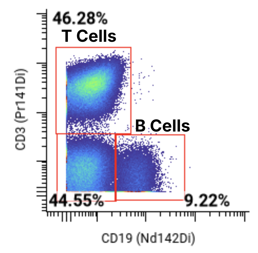
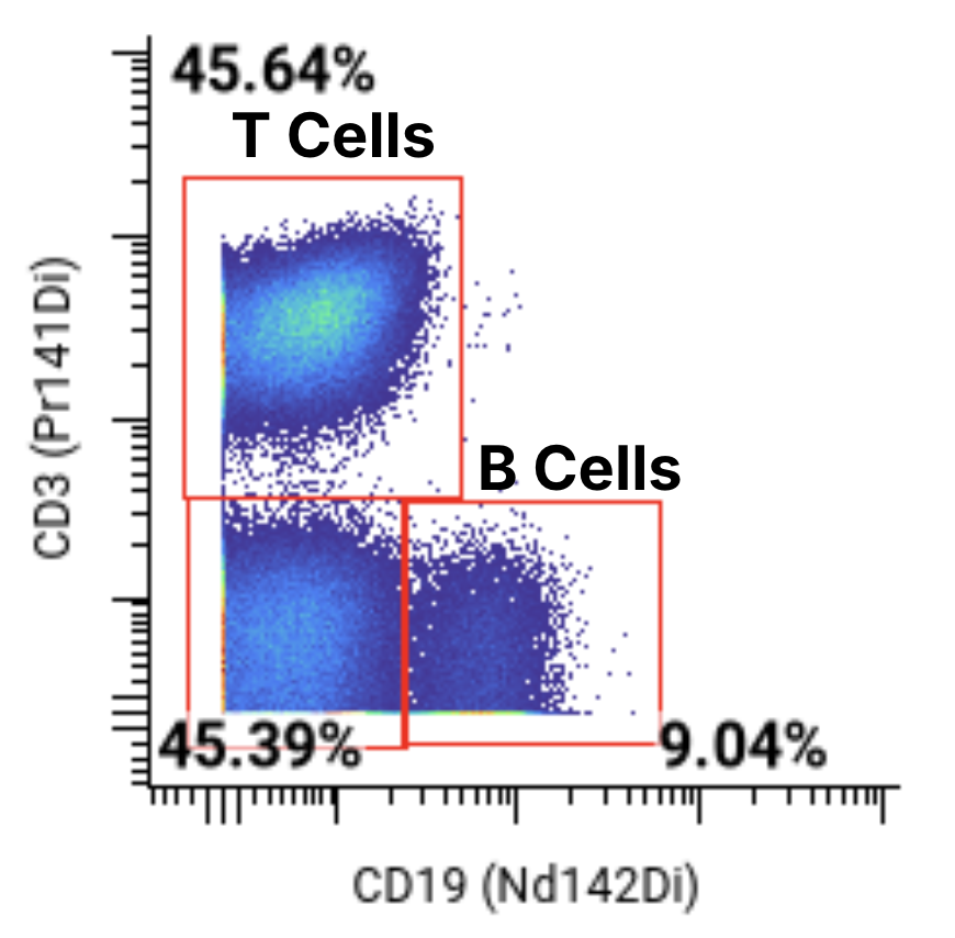
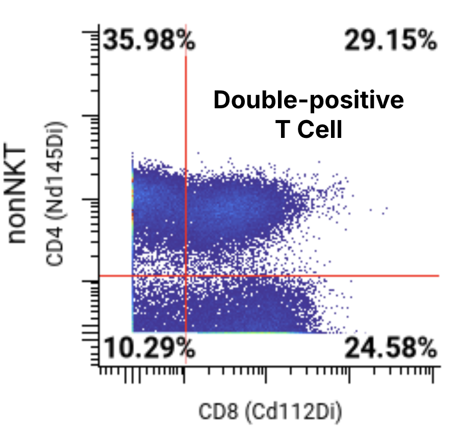
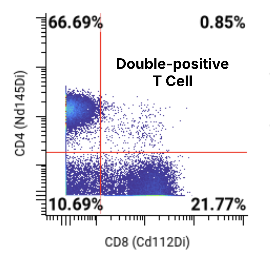
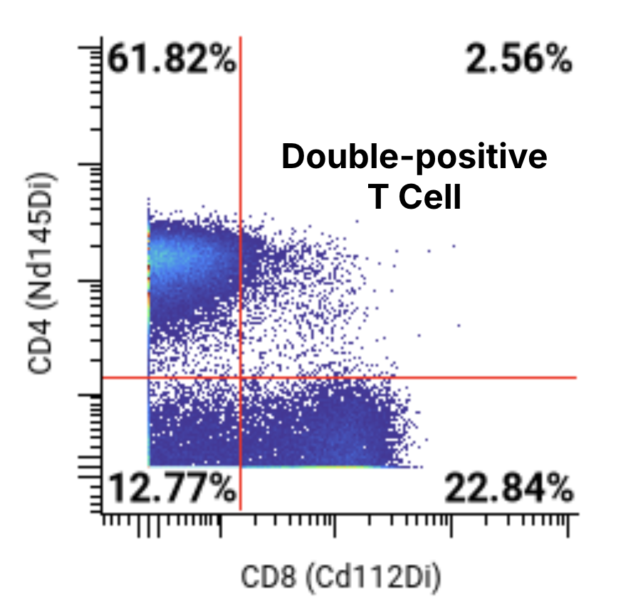
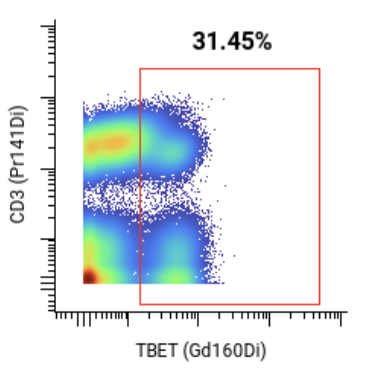
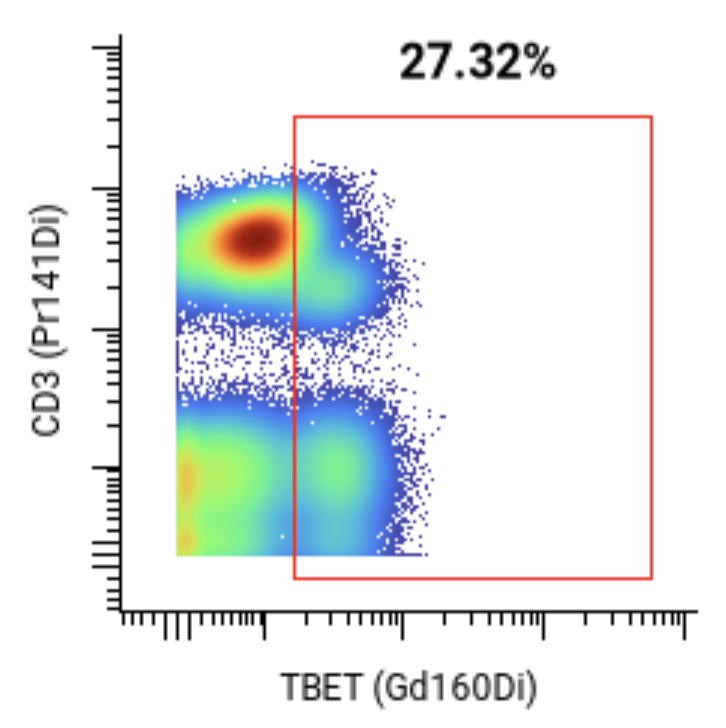
A 44-marker mass cytometry panel was applied to assess T, B, NK, myeloid, and other key immune cell populations

T cells
B cells
NK cells
Monocytes
Dendritic cells (DCs)
Neutrophils

Instrument

Cytometry analysis was performed on a CyTOF® Helios.

CytoChex had decreased B cell population frequencies.

Normally, B cells account for 5–10% of non-Granulocytes in healthy donors1. CytoChex resolved frequencies of 2-3% while Transfix and TokuKit preserved values within the expected range.
The reference range for B cells in healthy donors, when expressed as percent of non-granulocytes, is 5-10%. When evaluating B cell population frequencies:
  • Cytochex resolved a frequency of ~2-3% across sample conditions.
  • Transfix resolved a frequency of ~9-11% across sample conditions.
  • TokuKit resolved a frequency of ~7-9% across sample conditions.
CytoChex
Transfix
TokuKit

CytoChex had an increase in double-positive T cells after 7 days.

CD4⁺CD8⁺ T cells normally represent ~5–10% of total T cells in healthy donors2, but CytoChex samples reached ~30% after 7 days, a 3–6 fold increase.
The increase in CD4+CD8+ T cells suggests that CytoChex introduces artifacts after storing for greater than 7 days. When double-positive T cells were evaluated, as a percent of T cells:
  • CytoChex saw a frequency of about 30%.
  • Transfix saw a frequency of about 1%.
  • TokuKit saw a frequency of about 3%.
CytoChex
Transfix
TokuKit

Transfix did not show a clear T-bet+ population.

Transfix preserved a T-bet+ cell frequency of ~1%, compared to ~30% in CytoChex and TokuKit.
Based on Human Protein Atlas data, T-bet expression is expected in NK cells, γδ T cells, and subsets of memory T cells, corresponding to a fraction of both CD3+ and CD3- cells.3 However, in Transfix-preserved samples, a clearly defined T-bet+ population was not observed.
CytoChex
Transfix
TokuKit

Why does sample preservation  matter for your team?

Degradation distorts patient immune profiles
When preservation methods degrade or introduce artifacts, immune profiles shift, creating results that reflect preservation instability rather than a patient's true biology.
Failed samples waste time and budget
Degraded samples often fail QC, requiring costly redraws, repeat shipments, and delays in data delivery.
Inconsistencies undermine PD insights
Differences in how long samples sit before processing introduce noise, making it harder to detect real pharmacodynamic signals, mechanism of action, or biomarkers of response.

Want to learn more?

Ask us about the results of our CytoChex, Transfix, and TokuKit comparison—and what studies with TokuKit are coming next.
Thank you! Your submission has been received!
Oops! Something went wrong while submitting the form.

References

1 Miltenyi Biotec. MACS® handbook: Human cells and organs — Human cell sources: Blood (human) [Internet]. [cited 2025 Mar 3]. Available from: https://www.miltenyibiotec.com/US-en/support/macs-handbook/human-cells-and-organs/human-cell-sources/blood-human.html
2
González-Mancera MS, Bolaños NI, Salamanca M, Orjuela GA, Rodríguez AN, González JM. Percentages of CD4⁺CD8⁺ double-positive T lymphocytes in the peripheral blood of adults from a blood bank in Bogotá, Colombia. Turk J Haematol. 2020;37(1):36-41. doi:10.4274/tjh.galenos.2019.2019.0256
3 Huang C, Bi J. Expression regulation and function of T-Bet in NK cells. Front Immunol. 2021;12:761920. doi:10.3389/fimmu.2021.761920